Prescription Information
Understanding Prescriptions
- When a prescription is preceded by a;
+ This indicates Hypermetropic/far sighted vision , and when preceded by a
- This indicates Myopic/near sighted vision
R - Denotes Right eye. This is sometimes written as O.D, which is the abbreviated Latin equivalent.
L - Denotes Left eye. This is sometimes written as O.S, which is the abbreviated Latin equivalent. - Sphere (Sph): This figure represents the power in Dioptres of the spherical lens required to correct your sight. People who are Myopic or short sighted need negative powered lenses such as -1.00 or -3.00. The lenses change in 0.25 dioptre steps, myopic people can focus well for close work but cannot see distance clearly.
People who are Hypermetropic or long sighted need positive powered lenses such as +2.00 or +5.00. These lenses also change in 0.25 dioptre steps. Hypermetropic people generally have more difficulty seeing close work clearly however depending on the degree of Hypermetropia and age of the person the prescription may be required for distance viewing as well.
People who are Presbyopic or who just need glasses for reading also require positive powered lenses generally these people are over 40 years of age. These lenses enable the person to read but will blur the distance vision. - Cylinder (CYL) Toric Prescriptions: A Cylindrical lens or Cyl is the type of lens used to correct Astigmatism.
Astigmatism is caused by the eyes optical surfaces (such as cornea or lens of the eye) not being spherical, and hence being elliptical. This causes de-focusing of the eye for distance and near. To correct this problem a lens is required that has the power in only one direction or axis. This is a cylindrical lens. - Axis (Axis ) Toric Prescriptions: The axis relates the cylindrical lens, and denotes a reference point that indicates the direction of the power of the cylindrical lens. This is measured in degree steps from 1 degree to 180 degrees.
For Example :
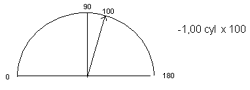
(The degree or orientation of prescription) - Prism: These lenses are used less frequently than sphere, Cyl, and ADD. Their function is to move an image up, down, left or right. This type of lens is used to correct double vision, either intermittent or constant (which is called a Tropia), or a tendency of the eyes to not work perfectly together, and one eye wanting to drift slightly, (this is called a Phoria).
Prism lenses are measured in units called Prism Dioptres . - Base: The Base relates to the direction of the Prism and denotes a reference point to tell us in which direction image has been moved. This direction will be recorded as either:
For Example: R � L (prism Dioptre) BASE IN � (prism Dioptre) BASE IN- Up
- Down
- In
- Out
- ADD (Add ) Bi-Focal Prescriptions The difference between the distance prescription and the reading prescription is known as the near addition, or ADD. This is only the case if a person is presbyopic and hence over 40 years of age. Presbyopic people either only need reading glasses or need a different prescription for reading than for distance.
For Example a distance prescription could be:
And a reading prescription could be:R
+1.00 sphere
� L
+1.00 sphere
R
+2.00 sphere
� L
+2.00 sphere
In this case the Add would be +1.00. As +1.00 Dioptres, needs to be added on to the distance prescription to create the reading prescription.
- Contact Lens Prescriptions - same as spectacle prescriptions only different A prescription for glasses is different than a prescription for contact lenses although they can be similar. A contact lens comes in direct contact with the cornea, which is the clear covering over the front surface of the eye. For this reason, there are additional measurements needed for fitting a contact lens and these need to be included in the prescription.
- The curvature of the cornea
- The diameter of the cornea
- Base Curve (BC) The curvature of the cornea is measured to determine the exact curvature (base curve) of the inside surface of the contact lens. This ensures the lens fits the shape of the eye providing optimum vision and comfort.
- Diameter (DIA) The diameter of the cornea is measured and also necessary to find the optimal contact lens diameter. This measurement is fairly straightforward. A contact lens is usually 2 - 4mm larger than the diameter of the cornea.
About the Eye
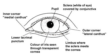
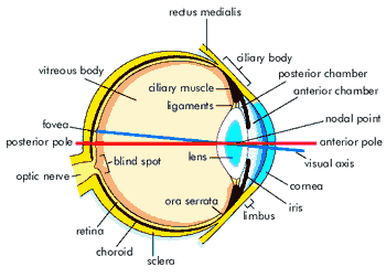
- The cornea is a curved, highly transparent tissue that separates air from clear fluid in the anterior chamber of the eye, which lies between the cornea and lens.
- The lens is a firm gel-like transparent tissue that is almost eight millimetres (one-third inch) in diameter and biconvex in shape, that is, thicker in the centre than at the edge. A thin transparent capsule surrounds the lens.
- The iris is in front of the lens and consists of a circular pigmented muscle that gives the eye its colour. The iris acts like the diaphragm of a camera and adjusts the amount of light that enters the eye through the hole in its centre called the pupil. Light then passes through the vitreous, a clear gel-like material that fills the centre of the eye, onto the retina.
- The retina is the film of the eye. It is a true extension of the brain and is composed of special nerve cells sensitive to light.
- The optic nerve is formed from these nerve cells and carries the light image entering the eye to the brain.
-
How the Eye Responds to Light An image on the retina stimulates photoreceptors, cells which, are specialised to detect light. This is a chemical event: pigments in the photoreceptors are broken down by light and this results in a generator potential. When threshold is reached, action potentials are created and nerve impulses begin to travel towards the brain.
The two photoreceptors in the human eye are the rods and cones. -
Rods: Rods contain a reddish-purple compound called rhodopsin, or visual purple. This contains opsin, a protein and retinene, a light-absorbing compound, which, is derived from vitamin A. When retinene is exposed to light, it changes shape (it is actually converted into an isomer) and this causes the rhodopsin to break down. The free opsin acts as an enzyme and, through a series of reactions, activates a neurotransmitter called cyclic GMP.
Cyclic GMP closes membrane pores in the rod cell and the negative charge inside the cell increases. This causes the membrane of the cell to, hyperpolarize, (become more negative) and sets up signals in nearby nerve cells. Nerve impulses then go the to brain for decoding. In the absence of further light stimulation the original retinene molecule reforms and this then recombines with opsin to form rhodopsin.
Rhodopsin is very sensitive to light and so rods are mainly used in dim light. In strong light the rhodopsin is broken down quicker than it can be reformed but in dim light, production is able to keep pace with the slower rate of breakdown. -
Cones: Cone cells contain the photosensitive pigment, iodopsin, a photosensitive pigment is made up of a photopsin, a different protein to that found in rods, and retinene. The events that occur in cone cells stimulated by light are basically the same as those that occur in rod cells. There are, however, three different types of cone. Each one contains a slightly different pigment and responds to a different wavelength of light. One responds to red light, one to green light and one to blue light, so allowing us to see in colour.
The colour that we 'see', or more accurately perceive, depends on which cones and stimulated. When all the cones are fully stimulated we see 'white'. When very few are stimulated we see black. Stimulation of separate types produces red, green or blue and all colours in between are produced by combinations of different levels of stimulation of the three types together.
Nearsightedness
If you can see objects nearby with no problem, but reading road signs or making out the writing on the board at school is more difficult, you may be near-or short-sighted.
The condition is called myopia, a term that comes from a Greek word meaning "closed eyes." Use of the word "myopia" for this condition may have grown out of one of the main indications of nearsightedness: Squinting to see distant objects clearly.
Myopia is not a disease. It simply refers to a variation in the shape of your eyeball. The degree of variation determines whether or not you will need corrective eyewear.
- What causes nearsightedness?
Myopia most often occurs because the eyeball is too long, rather than the normal, more rounded shape. Another less frequent cause of myopia is that the cornea, the eye's clear outer window, is too curved. There is some evidence that nearsightedness may also be caused by too much close vision work. The newest theory is that myopia is more commonplace in children that have had a night-light on when sleeping. This appeared to only cause myopia when the light was used on children under the age of two, after this age the � nightlight appears to have no effect on the development of myopia. 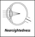 How does myopia affect sight?
Our ability to "see" starts when light enters the eye through the cornea. The shape of the cornea, lens and eyeball help bend (refract) light rays in such a manner that light is focused into a point precisely on the retina.
In contrast, if you are nearsighted, the light rays from a distant point are focused at a place in front of the retina. As the light will only be focused in that one place, by the time it reaches the retina it will have "defocused" again, forming a blurred image.- Who is affected by nearsightedness?
Myopia usually occurs between the ages of 8 to 12 years. Since the eyes continue to grow during childhood, near-sightedness almost always occurs before the age of 20. Often the degree of myopia increases as the body grows rapidly, and then levels off in adulthood.
During the years of rapid growth, frequent changes in prescription eyewear may be needed to maintain clear vision
Farsightedness
If you can see objects at a distance clearly but have trouble focusing well on objects close up, you may be farsighted.
Hypermetropia/Hyperopia. This causes the eyes to exert extra effort to see close up. After viewing nearby objects for an extended period, you may experience blurred vision, headaches and eyestrain. Children who are farsighted may find reading difficult.
Hypermetropia is not a disease. It simply means that you have a variation in the shape of your eyeball. The degree of variation will determine whether or not you will need corrective lenses.
- What causes farsightedness?
Hypermetropia most commonly occurs because the eyeball is too short; that is, shorter from front to back than is normal. The cornea having too little curvature may cause hypermetropia.
Exactly why eyeball shape varies is not known, but the tendency for farsightedness is inherited. Other factors may be involved too, but to a lesser degree than heredity. 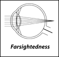 How does farsightedness affect sight?
Our ability to "see" starts when light enters the eye through the cornea. The shape of the cornea, lens and eyeball help bend (refract) light rays in such a manner that light is focused into a point precisely on the retina.
If, as in farsightedness, the eyeball is too short, the "point of light" focuses on a location behind the retina, instead of on the correct area of the retina, known as the fovea. As a result, at the point on the retina where a fine point of light should be focused, there is instead a disk-shaped area of light. Since light is not focused when it hits the retina, vision is blurred.
Convex lenses are prescribed to bend light rays more sharply and bring them to focus on the retina.- Who is affected by farsightedness?
Many people have a degree of farsightedness, yet it is only a problem if it significantly affects our ability to see well or causes headaches or eyestrain.
Astigmatism
If you experience a distortion or blurring of images at all distances -- nearby as well as far -- you may have astigmatism. Even if your vision is fairly sharp, headache, fatigue, squinting and eye discomfort or irritation may indicate a slight degree of astigmatism. A thorough eye examination, including tests of near vision, distant vision and vision clarity, can determine if astigmatism is present.
It means that you have a variation or disturbance in the shape of your cornea.
- What is astigmatism?
Astigmatism is one of a group of eye conditions known as refractive errors. Refractive errors cause a disturbance in the way that light rays are focused within the eye. Astigmatism often occurs with nearsightedness and farsightedness, conditions also resulting from refractive errors. - What causes astigmatism?
Astigmatism usually occurs when the front surface of the eye, the cornea, has an irregular curvature. Normally the cornea is smooth and equally curved in all directions and light entering the cornea is focused equally on all planes, or in all directions. In astigmatism, the front surface of the cornea is curved more in one direction than in the other. With the cornea's shape more like that of an American football or rugby ball than a basketball, the light hitting the more curved surface comes to a focus before that which enters the eye through the less curved surface. Thus, the light is focused clearly along one plane, but is blurred along the other so only part of anything being looked at can be in focus at any time.
This abnormality may result in vision that is much like looking into a distorted, wavy mirror. The distortion results because of an inability of the eye to focus light rays to a point. - Why are corneas shaped differently?
Not all corneas are perfectly curved, just as sets of teeth are seldom perfectly aligned. The degree of variation determines whether or not you will need corrective eyewear. If the corneal surface has a high degree of variation in its curvature, light refraction may be impaired to the degree that corrective lenses are needed to help focus light rays better.
The tendency to develop astigmatism is inherited. For that reason, some people are more prone to develop astigmatism than others. - How does astigmatism affect sight?
The crystal clear cornea is situated at the very front surface of the eye and enables light to enter the eyeball. The cornea accomplishes about four-fifths of the refractive work needed for clear vision, bending light rays toward one another into a point. The lens, located behind the cornea, further refines the refractive work begun by the cornea and directs the point of light toward a precise location on the retina, known as the fovea. If light is not focused into a fine point on the fovea, the image that reaches the retina cannot be clearly transmitted to the brain.
When astigmatism is present, the surface of the cornea is distorted instead of being spherical. It is unable to focus light rays entering the eye into the fine point needed for clear vision. At any time, only a small proportion of the rays are focused and the remainder are not, so that the image formed is always blurred. Usually, astigmatism causes blurred vision at all distances. - Who develops astigmatism?
Astigmatism is very common. Some experts believe that almost everyone has a degree of astigmatism, often from birth, which may remain the same throughout life.
Of interest to parents and those who work with children, astigmatism may contribute to poor schoolwork but is often not detected during routine eye screening in schools.
Presbyopia
Hold the book up close and the words appear blurred. Push the book farther away, and the words snap back into sharp focus.
That's how most of us first recognize a condition that eye care professionals call presbyopia, a name derived from Greek words meaning "old eye." Eye fatigue or headaches when doing close work, such as sewing, knitting or painting, are also common symptoms. Because it is associated with aging, presbyopia is often met with a groan -- and the realization that reading glasses or bifocals are inevitable.
- What causes presbyopia?
Recent research shows that the lens in the eye continues to grow as we age. Eventually it reaches a point, around the age of 45 that it is too big for the muscles that normally change it's shape to work effectively. The loss is gradual - but inevitable.
Our ability to "see" starts when light enters the eye through the cornea. The shape of the cornea, lens, and eyeball help bend (refract) light rays in such a manner that light is focused into a point precisely on the retina.
The crystalline lens plays a key role in focusing light on the retina. With the help of tiny ciliary muscles, it changes shape, or accommodates, for both near and distant objects by bending or flattening out to help focus light rays.� However, as one ages, not only does focusing on near objects become more difficult, the eye also is unable to adjust as quickly to rapid changes in focus on near and distant objects. - When does it occur?
The age at which presbyopia is first noticed varies, but it usually begins to interfere with near vision in the early 40's. - How is the problem diagnosed?
An accurate, thorough description of symptoms and a comprehensive eye health examination, including a testing of the quality of your near vision, are necessary to diagnose presbyopia. - How is presbyopia treated?
Usually, the treatment for presbyopia is prescription eyeglasses to help the eye accommodate for close-up work. Prescription lenses (reading glasses) help refract light rays more effectively to compensate for the loss of near vision.
If you do not have other vision problems, such as nearsightedness or astigmatism, you may only need glasses for reading or other tasks done at a close range. If you have other refractive errors, such as nearsightedness, bifocal or progressive addition lenses (in which the power of the lens changes gradually towards the bottom to allow reading, without the reading portion of the bifocal lens obviously visible) are often prescribed. - Can I still wear contact lenses?
Yes, you have three options with contact lenses: Bifocal contact lenses, monovision, and normal distance contact lenses with reading spectacles. Generally, bifocal contact lenses are not yet as successful as the normal "single vision" ones. - What lens option will work best for me?
Your eye care professional may ask a number of questions to help determine the best avenue of treatment. You may be asked to describe your usual lifestyle or daily activities and from this your practitioner will be able to recommend a solution most suited to your needs. For instance, if you are a librarian, your needs will be significantly different from those of a truck driver. - Once my vision is corrected for presbyopia, will I require frequent lens changes?
Presbyopia is a gradual change, happening over a number of years so your prescription will need to be updated periodically. Changes are best made at your regular eye examination rather than after the need for change starts to cause you difficulties.